The pain starts as a dull ache around or behind the kneecap – barely noticeable at first, then impossible to ignore. It gets worse going downstairs, worse after sitting for long periods, and worst of all during a run you have been training for for weeks. You have runner’s knee, and the instinct is to stop everything and rest.
Rest alone, however, is not the answer. Research consistently shows that complete rest from runner’s knee without targeted rehabilitation leads to recurrence rates above 70% – because the underlying biomechanical deficits that caused the condition remain unaddressed. The patella tracks abnormally, the hip abductors remain weak, the quadriceps are still imbalanced, and the first week back to running brings the pain right back.
The real solution is a specific, progressive exercise program that corrects the mechanical root causes of runner’s knee while the condition heals. This article gives you exactly that: the five most evidence-supported exercises for runner’s knee, explained in clinical detail – with the muscles targeted, the biomechanical rationale, the technique cues that make or break the movement, and the progression pathway from acute phase to return to running.
What Is Runner’s Knee? The Anatomy You Need to Understand
Runner’s knee is the colloquial term for patellofemoral pain syndrome (PFPS) – pain arising from abnormal contact pressure between the patella (kneecap) and the femoral trochlea (the groove in the femur where the patella sits). It is the most common running injury, accounting for approximately 25-40% of all running-related presentations to sports medicine clinics.
Why the Patella Tracks Abnormally
The patella does not sit passively in the trochlear groove – it is actively guided by the balance of forces acting on it. The quadriceps muscle – specifically the four heads of the quadriceps femoris (vastus medialis, vastus lateralis, vastus intermedius, and rectus femoris) – insert via the quadriceps tendon into the superior pole of the patella and collectively pull it superiorly during knee extension. The relative tension balance between the inner (vastus medialis oblique) and outer (vastus lateralis) heads determines whether the patella tracks centrally or laterally.
In runner’s knee, three contributing factors typically converge:
Vastus medialis oblique (VMO) weakness: The VMO – the teardrop-shaped inner quadriceps – fires during the last 30 degrees of knee extension to pull the patella medially. When the VMO is relatively weak compared to the vastus lateralis, the patella is pulled laterally with each step, creating abnormal contact pressure on the lateral facet of the patellofemoral joint.
Hip abductor and external rotator weakness: This is the most clinically underappreciated driver of runner’s knee. When the gluteus medius and external rotators are weak, the hip drops and internally rotates during single-leg loading (the “Trendelenburg pattern”) – which causes the femur to internally rotate and the knee to adduct (valgus collapse). This dynamic valgus dramatically increases the lateral tracking force on the patella, even when quadriceps balance is intact.
Iliotibial band (ITB) tightness: The ITB attaches to the lateral retinaculum – the fibrous tissue on the outer side of the patella. When tight, it pulls the patella laterally with each stride, amplifying the tracking abnormality and compounding the contact pressure problem.
The Clinical Consequence: Increased Patellofemoral Contact Pressure
The articular cartilage on the posterior surface of the patella has no nerve supply – which is why early patellofemoral degeneration is painless. The pain of runner’s knee arises from the richly innervated subchondral bone and the synovium (joint lining) that becomes inflamed when abnormal contact pressure is sustained. Peak patellofemoral contact pressure during running reaches 7-11 times body weight with each stride – a force that is normally distributed across the full contact area of the joint. When patellar malalignment concentrates this pressure on a smaller area, the result is the characteristic pain pattern of PFPS: anterior knee pain during loading, particularly with stairs, squatting, and running.
This mechanistic understanding is the direct rationale for every exercise in this article. If the problem is a force balance issue, the solution is restoring force balance through targeted strengthening and mobility work.
Before the Exercises: Two Essential Principles
Principle 1: Pain-Guided Progression
Every exercise in this program should be performed without exceeding a pain level of 3-4 out of 10. This is not a guideline to ignore pain entirely – it is a clinically validated threshold below which therapeutic loading of a tendon or joint structure promotes healing without causing further damage. Pain above 4/10 during an exercise indicates the load or range of motion is too provocative for the current tissue state. Reduce range, reduce resistance, or modify the movement rather than stopping entirely.
Principle 2: Proximal Before Distal
Research consistently shows that patients with runner’s knee who begin rehabilitation with hip strengthening before knee strengthening achieve faster pain reduction and return to sport than those who begin with isolated quadriceps work. This is because hip abductor and external rotator strength corrects the femoral internal rotation that drives patellar malalignment – addressing the upstream cause rather than the downstream consequence. The exercises in this article are sequenced accordingly: hip strength first, VMO and quadriceps second, mobility third.
Exercise 1: Side-Lying Hip Abduction
Primary muscles targeted: Gluteus medius, gluteus minimus, tensor fasciae latae Secondary muscles: Gluteus maximus, hip external rotators Clinical rationale: Directly strengthens the primary muscle responsible for frontal plane hip control during running, reducing dynamic knee valgus and the lateral patellar tracking force it generates
Why This Exercise Is the Foundation of Runner’s Knee Rehabilitation
A meta-analysis published in BMC Musculoskeletal Disorders found that hip abductor strengthening produced significant improvements in knee pain and functional outcomes across multiple lower limb conditions – including patellofemoral pain syndrome. The mechanism is straightforward: during each single-leg stance phase of running, the gluteus medius must generate sufficient force to prevent the pelvis from dropping on the non-stance side. When it cannot, compensatory femoral internal rotation occurs at the knee – driving the valgus collapse that laterally displaces the patella.
Technique
- Lie on your side with your hips stacked directly above each other – not rolled forward or backward
- Both legs are straight or the bottom knee slightly bent for stability
- Keep the top foot dorsiflexed (toes toward shin) to ensure the lift comes from the hip, not the ankle
- Lift the top leg to approximately 30-45 degrees above horizontal – do not lift higher, as this recruits the hip flexors rather than the abductors
- Pause for 2 seconds at the top of the movement
- Lower slowly over 3-4 seconds – the eccentric phase is where the most significant strength adaptation occurs
- Keep the top hip from rolling backward – the tendency to rotate the hip externally is a compensation pattern that reduces gluteus medius activation
Common errors to avoid:
- Rotating the trunk backward to “cheat” the lift – the pelvis must stay perpendicular to the floor throughout
- Pointing the toes toward the ceiling – this shifts the load to the TFL and reduces gluteus medius isolation
- Lifting too high – this causes hip flexor substitution
Dosage: 3 sets of 15-20 repetitions each side. Progress by adding a resistance band just above the knee when 20 repetitions feel comfortable. Ultimate progression: standing resisted hip abduction on a cable machine, which more closely replicates the functional demand of single-leg stance during running.
Exercise 2: Clamshell
Primary muscles targeted: Gluteus medius (posterior fibers), piriformis, obturator internus Secondary muscles: Gluteus maximus, hip external rotators Clinical rationale: Specifically activates the posterior hip external rotators that resist femoral internal rotation – the primary driver of dynamic knee valgus in runners
The Clamshell’s Specific Contribution
While side-lying hip abduction targets the superior and middle fibers of the gluteus medius, the clamshell selectively recruits the posterior fibers and the deep external rotators (piriformis, obturator internus, gemelli) that are particularly responsible for resisting the internal rotation that occurs at impact during running. Electromyography studies confirm that the clamshell produces high gluteus medius and posterior hip rotator activation with minimal TFL recruitment – making it the most specific exercise available for correcting the rotation pattern that drives runner’s knee.
Technique
- Lie on your side with your hips at 45-60 degrees of flexion and knees at 90 degrees of flexion – this hip flexion angle is critical for isolating the gluteus medius and external rotators, rather than the hip abductors in a neutral position
- Stack your hips directly above each other and keep your feet together throughout the movement
- Rotate the top knee upward – like a clamshell opening – as far as possible without the pelvis rotating backward
- The key movement cue: imagine a ball in your back pocket that must not fall out – this prevents the pelvis from rolling backward as the hip opens
- Hold the open position for 2 seconds, squeezing the glute
- Return slowly over 3-4 seconds
- The feet stay together throughout – if they separate, the movement is being performed incorrectly
Resistance progression: Begin with body weight. Add a resistance band just above the knees when 20 repetitions feel easy. Advance to side-lying with a resistance band and hip at neutral (not flexed) to target the abductors in a more functional position.
Dosage: 3 sets of 15-20 repetitions each side. Perform before hip abduction in the session, as its more specific external rotator recruitment prepares the neuromuscular pattern for the abduction work.
Find out more about how hip abductor weakness drives knee pain and joint loading – the biomechanics of why proximal hip strength is central to knee health is covered in the context of joint-protective exercise programming.
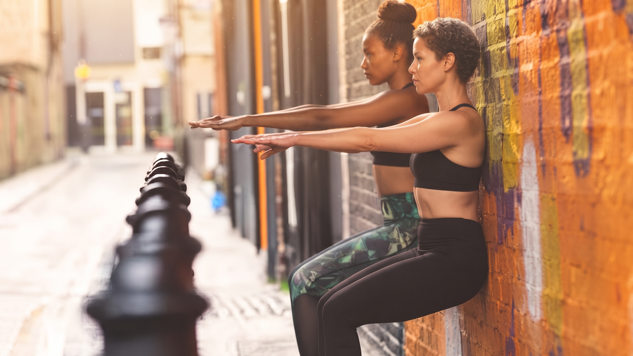
Exercise 3: Wall Sit with VMO Emphasis
Primary muscles targeted: Vastus medialis oblique (VMO), quadriceps femoris, gluteus maximus Secondary muscles: Soleus, hip external rotators Clinical rationale: Strengthens the VMO in an isometric and eccentric position that reduces patellofemoral contact pressure compared to traditional squats, while creating sufficient neuromuscular stimulus for VMO hypertrophy
Why Not a Regular Squat?
Traditional deep squats significantly increase patellofemoral contact pressure as knee flexion increases beyond 60 degrees – the opposite of what the recovering knee needs. Patellofemoral contact area and pressure are minimized between 0-45 degrees of knee flexion and increase substantially beyond this range. The wall sit performed at 45-60 degrees of knee flexion allows meaningful quadriceps and VMO loading while keeping contact pressure within a tolerable range for the irritated patellofemoral joint.
The wall sit also has an important advantage over dynamic squats: its isometric nature eliminates the moving contact point of dynamic patellar tracking, allowing VMO to strengthen without the cyclical irritation of repeated loading and unloading across a painful arc.
Technique
- Stand with your back flat against a wall and your feet hip-width apart, approximately 30-45cm away from the wall
- Slide your back down the wall until your knees are at approximately 45-60 degrees of flexion – for most people this means thighs are somewhere between parallel and 45 degrees above horizontal
- Ensure your knees track directly over your second toe – not caving inward (valgus) and not bowing outward
- Press your lower back flat into the wall – anterior pelvic tilt during a wall sit shifts load away from the quadriceps and onto the lumbar spine
- Hold the position for 20-30 seconds initially, breathing consistently
- For VMO emphasis: place a ball or yoga block between the thighs just above the knees and gently squeeze it isometrically throughout the hold – this activates hip adductors which have a synergistic relationship with VMO activation
- Slide back up slowly
Progression pathway:
- Phase 1: 3 x 20-30 second holds at 45 degrees
- Phase 2: 3 x 30-45 second holds at 50-55 degrees
- Phase 3: 3 x 45-60 second holds at 60 degrees
- Phase 4: Single-leg wall sit (one leg extended forward) – dramatically increases VMO demand
Warning: If pain increases above 4/10 during the wall sit, reduce the knee flexion angle (come higher up the wall) rather than stopping entirely.
Exercise 4: Step-Down (Eccentric Knee Control)
Primary muscles targeted: Quadriceps (eccentric), gluteus medius, gluteus maximus Secondary muscles: Hip external rotators, tibialis anterior Clinical rationale: The step-down is the single most functionally relevant exercise for runner’s knee because it replicates the eccentric quadriceps demand of descending stairs and downhill running – the exact motion that is most painful in PFPS – in a controlled, progressable format
The Eccentric Principle in Runner’s Knee Rehabilitation
Eccentric strengthening – where the muscle generates force while lengthening – has consistently demonstrated superior efficacy to concentric-only training for patellofemoral pain rehabilitation. During downhill running and stair descent, the quadriceps must control the knee’s flexion against gravity – an eccentric contraction. Weakness or poor neuromuscular control of this eccentric phase is what produces the sharp catching pain that patients with runner’s knee experience going downstairs – and training this specific demand is what resolves it.
The step-down also uniquely reveals dynamic knee alignment in a functional context. If the knee caves inward (valgus) during the step-down, it exposes the hip abductor and external rotator weakness that needs to be addressed before progressing to running.
Technique
- Stand on a step (approximately 10-15cm high initially) on the affected leg, with the other foot hanging free beside it
- Arms out to the sides for balance
- Slowly lower the free foot toward the floor by bending the stance knee to 30-40 degrees
- The pace of the lowering phase should take 3-4 seconds – this slow eccentric controls the neuromuscular demand
- Critical alignment check: throughout the movement, the stance knee should track directly over the second toe – not diving inward. If valgus occurs, the hip abductors are insufficient for this level of demand; return to the hip exercises above
- Touch the heel to the floor lightly – do not transfer weight – and return to the starting position
- The ascending phase should also be controlled (not a sudden push-up)
The knee alignment principle: If you observe the knee diving inward during the step-down, this is the most important biomechanical finding you can get. It tells you definitively that hip abductor and external rotator strength – not quadriceps strength – is the primary limiting factor for this patient. Return emphasis to Exercises 1 and 2 before progressing.
Progression:
- Phase 1: Step-down from 10cm step, 30 degrees knee flexion, 3 x 10 repetitions
- Phase 2: Step-down from 15cm step, 40 degrees flexion, 3 x 15 repetitions
- Phase 3: Step-down from 20cm step, 50 degrees flexion, 3 x 15 repetitions
- Phase 4: Add 5-10% body weight in a light backpack for additional eccentric loading
Dosage: 3 sets of 10-15 repetitions. Quality of movement is paramount – perform only as many repetitions as you can maintain perfect knee alignment.
Find out more about managing osteoarthritis naturally with exercises that prioritize knee alignment and progressive loading – the same alignment principles that govern runner’s knee rehabilitation apply to degenerative knee conditions.
Exercise 5: Standing ITB and Hip Flexor Mobility
Areas targeted: Iliotibial band, hip flexors (iliopsoas, rectus femoris), lateral quadriceps Clinical rationale: Reduces the lateral retinacular tension that contributes to lateral patellar displacement, and addresses the hip flexor tightness that increases anterior pelvic tilt and alters patellofemoral mechanics during running
Why Mobility Is the Final Piece
Strengthening the hip abductors and VMO corrects the active force imbalances driving runner’s knee. But even a strong hip cannot fully compensate for a tight ITB that is mechanically pulling the patella laterally with every stride. Studies confirm that ITB tightness measured by the Ober test is significantly associated with patellofemoral pain syndrome, and that addressing it alongside strengthening produces faster pain resolution than strengthening alone.
Hip flexor tightness is an equally important but less discussed contributor. When the iliopsoas and rectus femoris are shortened, they create an anterior pelvic tilt that increases lumbar lordosis and, through the kinetic chain, alters the angle at which the quadriceps pull on the patella – increasing the Q-angle and the lateral vector force on the kneecap.
Part A: Standing ITB Stretch (Cross-Leg Lateral Lean)
- Stand near a wall for balance if needed
- Cross the affected leg behind the non-affected leg
- Push the affected hip out laterally to the side while leaning the upper body away from the affected side
- You should feel a stretch along the outer thigh and hip of the affected side
- Hold for 30-45 seconds
- The key technique point: keep both feet on the floor and the pelvis neutral – do not hike the hip upward, as this changes the stretch to the lumbar spine rather than the ITB
- Repeat 3 times on each side
Important clinical note: Foam rolling the ITB before this stretch enhances its effectiveness by reducing myofascial tension that limits the stretch. Roll slowly from the knee to the hip along the lateral thigh, spending additional time on particularly tender points. Foam rolling the lateral quadriceps and ITB reduces tissue tension and temporary increases extensibility, allowing the subsequent stretch to work at greater range. Do not foam roll over the lateral knee itself – this compresses the ITB over the lateral femoral condyle, the exact area of most runners’ ITB friction syndrome.
Part B: Kneeling Hip Flexor Stretch
- Kneel on the affected knee with the foot behind (kneeling lunge position)
- The non-affected leg is in front with foot flat on the floor
- Keeping your torso upright, push the pelvis forward and slightly downward – you should feel a stretch in the front of the hip and thigh of the kneeling leg
- Add a posterior pelvic tilt (tuck the pelvis under) to deepen the stretch on the rectus femoris specifically – the component most relevant to patellofemoral mechanics
- Hold for 30-45 seconds
- Advance by reaching the ipsilateral arm (same side as the kneeling leg) overhead and laterally – this adds a lateral trunk lean that further elongates the iliopsoas
- Repeat 3 times each side
Frequency: Both stretches in Exercise 5 should be performed daily – morning and evening when symptomatic. Unlike strength exercises that require recovery time, flexibility work benefits from frequent low-intensity repetition.
Find out more about yoga for bone and joint health – yoga-based hip mobility sequences that incorporate similar hip flexor and lateral chain stretching in a progressive, functional context.
The Complete Runner’s Knee Rehabilitation Schedule
Acute Phase (Weeks 1-2): Pain Reduction and Foundation
During this phase, running is restricted or replaced with pain-free cross-training (swimming, cycling, elliptical). The goal is pain reduction and establishing the neuromuscular foundation for subsequent loading.
Daily:
- Exercise 5 (ITB stretch + hip flexor stretch): Morning and evening
- Ice to the anterior knee: 15-20 minutes after any activity
3 days per week:
- Exercise 1 (side-lying hip abduction): 3 x 15 each side
- Exercise 2 (clamshell): 3 x 15 each side
- Exercise 3 (wall sit): 3 x 20 seconds at 45 degrees
Cross-training: Stationary cycling (seat elevated to reduce knee flexion), swimming, or elliptical – maintaining cardiovascular fitness without patellofemoral loading. Find out more about joint-protective cross-training during injury recovery and which modalities achieve running-equivalent cardiovascular stimulus without knee impact.
Strengthening Phase (Weeks 3-5): Building Capacity
Pain should be consistently below 3/10 before progressing to this phase. Running may be reintroduced at 25-30% of normal volume on flat surfaces only.
4 days per week:
- Exercise 1 (hip abduction with resistance band): 3 x 20
- Exercise 2 (clamshell with resistance band): 3 x 20
- Exercise 3 (wall sit): 3 x 45 seconds at 55 degrees
- Exercise 4 (step-down from 15cm): 3 x 12
Daily: Exercise 5 mobility work
Return to Running Phase (Weeks 6-8): Progressive Loading
Full running volume is reintroduced at 10% per week. All five exercises continue at maintenance frequency (3x/week), with progression to single-leg loading.
3 days per week:
- Exercise 1 (standing cable hip abduction): 3 x 15
- Exercise 2 (resistance band clamshell, 30-second holds): 3 sets
- Exercise 3 (single-leg wall sit): 3 x 20 seconds each leg
- Exercise 4 (step-down from 20cm with backpack): 3 x 15
Running reintroduction: Start with flat, soft surface runs at reduced pace. Avoid downhill running until the step-down can be performed with perfect knee alignment at 20cm height without pain.
Twice daily: Exercise 5 mobility – continuing indefinitely as maintenance
Nutrition and Botanical Support for Runner’s Knee Recovery
The exercises rebuild the strength and biomechanical foundation. Targeted nutritional and botanical support reduces the inflammatory burden and provides the molecular building blocks for cartilage and connective tissue repair.
Collagen Peptides: Rebuilding the Joint Matrix
Hydrolyzed collagen peptides consumed 30-60 minutes before exercise have been shown to increase collagen synthesis in joint tissue – a particularly relevant finding for PFPS, where the articular cartilage on the posterior patellar surface is subjected to abnormal contact pressure with each stride. The Naturem™ Joints+ formula delivers hydrolyzed crocodile bone collagen peptides processed through a patented hydrolysis method for maximum bioavailability, combined with Drynaria Fortunei for osteoblast and connective tissue stimulation, and Rhizoma Homalomena for microcirculation enhancement that improves nutrient delivery to the poorly vascularized articular cartilage. Find out more about the joint health supplement evidence base and how each ingredient in Naturem™ Joints+ addresses a specific component of joint degeneration and repair.
Omega-3 Fatty Acids: Reducing the Inflammatory Response
The synovial inflammation of acute runner’s knee is driven by prostaglandins and IL-1β – the same mediators that omega-3 EPA and DHA directly modulate. Supplementation at 2-3g EPA+DHA daily reduces inflammatory cytokine production and has been shown to reduce exercise-induced joint inflammation in multiple clinical trials. This anti-inflammatory support is particularly valuable in the acute phase of runner’s knee when the irritated synovium is driving the pain response.
Anti-Inflammatory Diet: The Systemic Context
Chronic low-grade systemic inflammation amplifies the local joint inflammation of runner’s knee through the NF-kB pathway that drives IL-6 and TNF-alpha production. A Mediterranean-style dietary pattern – emphasizing fish, olive oil, colorful vegetables, legumes, and whole grains while minimizing ultra-processed foods – reduces this systemic inflammatory substrate and creates the biochemical environment most conducive to tissue repair.
When to See a Clinician: Red Flags and Diagnostic Considerations
The five exercises above are appropriate for most cases of patellofemoral pain syndrome – the most common diagnosis in runners presenting with anterior knee pain. However, several conditions present similarly to runner’s knee but require different management:
Patellar tendinopathy (jumper’s knee): Pain is localized to the inferior pole of the patella (the tendon attachment), not diffusely anterior. Wall sits and step-downs may aggravate it differently than PFPS. Requires tendon-loading protocols rather than patellofemoral offloading.
Meniscal pathology: Typically presents with medial or lateral joint line pain rather than anterior kneecap pain, often with a mechanical catching or locking sensation. Requires imaging and specialist evaluation before exercise prescription.
Plica syndrome: A thickened band of synovium that snaps over the medial femoral condyle during knee flexion – produces a reproducible clicking and medial knee pain that worsens with squatting. Often misdiagnosed as PFPS.
Seek medical evaluation if:
- Knee swelling is present (suggests intra-articular pathology – meniscal tear, cartilage damage, synovitis)
- Pain does not improve with 3-4 weeks of the rehabilitation program
- Pain is located medially or laterally on the joint line rather than anteriorly
- You have a history of knee instability or previous significant trauma
- Any locking or catching sensation occurs during movement
Find out more about joint health and when to seek specialist assessment – distinguishing between the various knee conditions that require different management approaches is essential for choosing the right rehabilitation strategy.
Traditional Medicine Perspectives on Knee Pain in Runners
In Traditional Chinese Medicine (TCM), the knee is governed primarily by the Liver and Kidney meridians – the Liver governing the sinews (tendons and ligaments) and the Kidney governing bone integrity and the fundamental constitutional reserve. Runner’s knee, in TCM terms, arises from a combination of “strenuous exertion consuming Liver Blood” (the Blood that nourishes the sinews becoming insufficient with excessive running volume) and “Wind-Cold-Damp obstructing the knee channel” (the local inflammatory obstruction that produces pain and stiffness).
Treatment focuses on two simultaneous therapeutic goals: tonifying Liver Blood and Kidney essence to restore the nutritive foundation of tendon and bone health, and clearing the local obstruction through improved Qi and Blood circulation to the knee. The herbs used for this pattern – Drynaria Fortunei (Gu Sui Bu, Kidney yang tonic that strengthens sinew and bone), Rhizoma Homalomena (Qian Nian Jian, “thousand year health” – a classic joint and sinew herb) – are precisely those now confirmed by modern pharmacology to have osteoblast-stimulating, anti-inflammatory, and microcirculatory effects relevant to patellofemoral rehabilitation.
In Vietnamese traditional medicine (Y học cổ truyền), the equivalent concept of “đau gối do gân cốt hư” – knee pain from sinew and bone deficiency – reflects the same understanding that the knee’s health depends on the systemic constitutional vitality of the Kidney and Liver organ systems. Traditional remedies include herbs that both locally relieve pain and inflammation and systemically nourish the structures affected. This integrated approach – addressing local biomechanics through exercise and systemic connective tissue health through nutrition and botanical support – represents the most comprehensive model available for runner’s knee rehabilitation.
Conclusion: Runner’s Knee Is a Biomechanical Problem With a Biomechanical Solution
Runner’s knee did not develop because you ran – it developed because you ran with insufficient hip abductor strength, inadequate VMO activation relative to your vastus lateralis, and either tight ITB or limited hip flexor flexibility. Each of those contributing factors is entirely addressable through the exercises in this article.
The five-movement program – clamshell, side-lying hip abduction, wall sit, step-down, and ITB/hip flexor mobility – addresses every link in the biomechanical chain that produces patellofemoral pain, sequenced to progress logically from proximal hip control through to eccentric knee loading and flexibility work. Add the nutritional and botanical support to reduce the inflammatory burden and supply the molecular building blocks for connective tissue repair, and you have the most comprehensive rehabilitation approach current evidence supports.
Your running will come back. Follow the program, respect the pain-guided progression, and the patella will track correctly again.
This article is for educational purposes only and does not constitute individual medical advice. If your knee pain is severe, accompanied by swelling, or does not improve with 3-4 weeks of rehabilitation, please seek assessment from a physiotherapist or sports medicine physician.
Frequently Asked Questions (FAQs)
With consistent adherence to the rehabilitation program described, most cases of runner’s knee show significant pain reduction within 4-6 weeks and return to full running capacity within 8-12 weeks. The critical factor is patient compliance: research shows that supervised rehabilitation with three or more sessions per week produces significantly faster outcomes than unsupervised home exercise alone. Long-standing runner’s knee (symptoms present for more than 3 months) typically requires 12-16 weeks of consistent rehabilitation. (NCBI, 2017)
In the acute phase (pain above 4/10 during running), running should be replaced with low-impact cross-training. Once pain is consistently below 3/10 at rest and during activities of daily living, walking and light jogging on flat surfaces can be reintroduced at 25-30% of your previous volume, increasing by no more than 10% per week. Avoiding downhill running until the step-down exercise can be performed at 20cm height with perfect knee alignment is one of the most clinically important return-to-running criteria for PFPS. (NCBI, 2017)
The step-down (Exercise 4) is the single most functionally specific exercise for runner’s knee – it replicates the exact eccentric quadriceps demand of the most provocative activities (stairs, downhill running) while simultaneously revealing hip abductor weakness through the dynamic valgus pattern it exposes. However, if the knee valgus during step-down is severe, beginning with the clamshell (Exercise 2) and side-lying hip abduction (Exercise 1) for 2-4 weeks first will make the step-down significantly safer and more effective. (NCBI, 2022)
Yes – McConnell patellar taping, which manually repositions the patella medially using adhesive tape, has good evidence for reducing pain immediately and allowing more comfortable participation in rehabilitation exercises during the acute phase. Kinesiology tape applied in a similar medial direction has comparable evidence. Taping does not correct the underlying weakness – it is a pain management adjunct that allows you to exercise with better form while the strengthening produces lasting correction. (NCBI, 2017)
Evidence for collagen peptides (10-15g daily, taken with vitamin C before exercise or rehabilitation) in supporting cartilage matrix repair and tendon collagen synthesis is mechanistically credible and supported by emerging clinical evidence. Naturem™ Joints+ combines hydrolyzed collagen peptides with anti-inflammatory botanicals (Clinacanthus Nutans, Rhizoma Homalomena) and bone/tendon support herbs (Drynaria Fortunei) in a formula that addresses both the structural and inflammatory dimensions of patellofemoral recovery. It is a complement to – not a substitute for – the strengthening and mobility work described in this article. (Naturem, 2026)
References:
Crossley, K. M., van Middelkoop, M., Barton, C. J., & Culvenor, A. G. (2019). Rethinking patellofemoral pain: Prevention, management and long-term consequences. Best Practice and Research Clinical Rheumatology, 33(1), 48-65. https://doi.org/10.1016/j.berh.2019.02.004
Thomas, D. T., Shruthi, R., Prabhakar, A. J., Dineshbhai, P. V., & Eapen, C. (2022). Hip abductor strengthening in patients diagnosed with knee osteoarthritis: A systematic review and meta-analysis. BMC Musculoskeletal Disorders, 23, 576. https://doi.org/10.1186/s12891-022-05557-6
Lack, S., Barton, C., Sohan, O., Crossley, K., & Morrissey, D. (2015). Proximal muscle rehabilitation is effective for patellofemoral pain: A systematic review with meta-analysis. British Journal of Sports Medicine, 49(21), 1365-1376.
Neal, B. S., Barton, C. J., Gallie, R., O’Halloran, P., & Morrissey, D. (2016). Runners with patellofemoral pain have altered biomechanics which targeted interventions can modify: A systematic review and meta-analysis. Gait and Posture, 45, 69-82.
Nascimento, L. R., Teixeira-Salmela, L. F., Souza, R. B., & Resende, R. A. (2018). Hip and knee strengthening is more effective than knee strengthening alone for reducing knee pain and improving function in individuals with patellofemoral pain. Journal of Orthopaedic and Sports Physical Therapy, 48(1), 19-31.