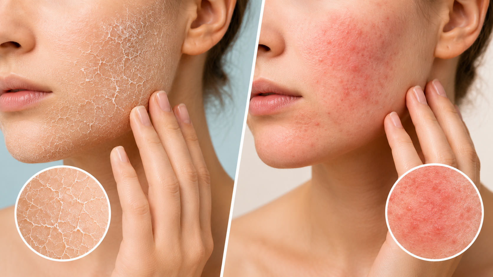
Your skin feels tight, rough, and uncomfortable. It itches. It flakes. You reach for a moisturizer – and it helps a little, or perhaps not at all.
Is this dry skin? Or is it something more?
This question matters more than most people realize. Dry skin and eczema are two conditions that overlap significantly in appearance but are fundamentally different in origin, severity, and treatment. Treating eczema like ordinary dry skin can leave the condition unmanaged for years – allowing it to worsen, spreading to new areas, and potentially contributing to the development of asthma and allergic conditions through a process called the atopic march.
Eczema affects approximately 31 million Americans – about 12% of children and 7% of adults in the United States. It is one of the most common skin conditions in the world – yet it is also one of the most commonly misidentified, mistaken for ordinary environmental dryness by patients and sometimes even clinicians.
This article gives you the clinical knowledge to tell the difference – accurately, confidently, and with an understanding of what is actually happening beneath the surface of your skin.
The Fundamental Difference – What Each Condition Actually Is
What Dry Skin Is
Dry skin – clinically called xerosis – is a condition in which the skin lacks adequate moisture in the outer layer (stratum corneum). It is not a disease. It is a common, usually temporary disruption of the skin’s moisture balance driven by external factors.
Common external causes of dry skin include:
- Cold or dry climates that reduce ambient humidity
- Indoor heating during winter months – significantly lowers indoor humidity
- Long, hot showers that strip natural skin oils
- Frequent hand washing with harsh soaps
- Chemical irritants in detergents, cleaning products, and skincare
- Aging – skin naturally becomes thinner and drier with age
Dry skin is extremely common – it affects people of all ages, and skin becomes progressively drier and thinner as we age. When the external cause is addressed, dry skin typically resolves with appropriate moisturization and gentle skincare. It does not involve immune system dysfunction. It does not cause systemic consequences. It does not progress.
What Eczema Is
Eczema – most commonly atopic dermatitis (AD) – is a fundamentally different condition. It is a chronic inflammatory skin disease driven by a combination of genetic predisposition, immune system dysregulation, skin barrier dysfunction, and environmental triggers.
Eczema happens when the outer skin barrier layer does not work as it should. Moisture escapes. Allergens, irritants, and pathogens penetrate more easily. The immune system – already dysregulated – overreacts to these penetrating substances and triggers chronic inflammation.
The key distinction: dry skin is a moisture problem. Eczema is an immune and barrier problem – with dryness as one of its consequences.
A combination of genes, a faulty immune response, and environmental triggers cause eczema. There is a strong hereditary component – it runs in families, often alongside asthma and hay fever. The condition is not curable, though it is highly manageable with the right approach.
The Biology Behind Eczema – Why It Is So Much More Than Dryness
Understanding why eczema is a disease – not just a skin care problem – requires a brief look at what is happening beneath the surface.
The Filaggrin Failure
Filaggrin is a structural skin protein that plays a central role in maintaining the epidermal barrier. It binds keratin fibers together to form the tough outer layer of skin and breaks down into natural moisturizing factors that keep skin hydrated and at the correct pH.
In people with eczema, mutations in the filaggrin gene – FLG – are among the strongest genetic risk factors for the condition. When filaggrin is deficient or dysfunctional, the skin barrier is structurally compromised at a molecular level. Transepidermal water loss (TEWL) increases significantly – the skin dries out rapidly. Allergens and pathogens penetrate more deeply. And the immune system in genetically predisposed individuals mounts an abnormal response.
This is not dryness that moisturizer alone can fix. It is a structural and immunological defect that no amount of topical hydration fully corrects.
The Immune Dysregulation – Th2 Dominance
Eczema is primarily driven by a Th2-dominant immune response – an overactivation of the immune pathways associated with allergy and inflammation. Elevated levels of interleukins IL-4, IL-13, and IL-31 drive allergic inflammation and the intense, relentless itch that characterizes the condition.
IL-31 in particular is directly responsible for the neurogenic itch signaling in eczema. The hallmark intense itching of eczema – which often precedes the visible rash – is a direct consequence of this cytokine activity rather than simply skin dryness.
The Gut-Skin Axis
Emerging research is revealing that the gut microbiome plays a significant and bidirectional role in eczema development and severity. Gut microbiota alterations disrupt skin barrier homeostasis through the gut-skin axis. Microbial imbalance reduces filaggrin expression, increases skin permeability, and triggers inflammation. Short-chain fatty acids (SCFAs) produced by beneficial gut bacteria reduce skin inflammation and enhance skin barrier function by promoting regulatory T cell activity and suppressing pro-inflammatory cytokines IL-6 and TNF-α.
This gut-skin connection explains why dietary interventions and gut health approaches can measurably influence eczema outcomes – and underscores why eczema is a systemic condition rather than a localized skin problem. For more on how gut health connects to systemic immune function, find out more in Naturem’s anti-inflammatory diet guide.
The 7 Key Signs That Tell Dry Skin and Eczema Apart
Sign 1 – The Quality and Location of Itching
Both conditions itch – but the character of that itch is profoundly different.
Dry skin causes mild to moderate itching that is typically localized to the dry area, comes on gradually, and is significantly relieved by moisturizing. It is an itch that is proportionate to the visible skin change.
Eczema causes intense, often unbearable itching that can occur even before any visible rash appears. In fact, eczema is classically described as “the itch that rashes” – the itch comes first, the rash results from scratching. The itching is often worst at night, disrupting sleep. It is frequently described as burning, deep, and impossible to satisfy with scratching. Moisturizer may provide minimal or no relief for eczema itch – because the itch is neurogenic and immune-driven, not simply surface dryness.
Clinical rule: If moisturizing consistently relieves the itch – likely dry skin. If the itch persists, worsens at night, or precedes visible skin changes – suspect eczema.
Sign 2 – The Nature of the Rash and Skin Changes
Dry skin presents as rough, flaky texture, mild tightness, and a dull, ashy appearance. The scaling is fine and diffuse. There is typically no prominent redness, no defined borders, and no raised lesions.
Eczema presents with a distinct rash pattern that includes:
- Redness and inflammation – actively inflamed patches, often with warm-to-touch skin
- Raised, thickened plaques – the skin becomes leather-like in chronic eczema through a process called lichenification
- Small fluid-filled blisters – particularly in dyshidrotic eczema affecting the hands and feet
- Weeping and oozing – the rash can weep clear or yellowish fluid, particularly when infected
- Crusting – dried weeping fluid forms crusts on the skin surface
- Defined borders – eczema patches often have reasonably distinct margins
On darker skin tones, eczema may appear as darkened, greyish, or purplish patches rather than the red appearance typical on lighter skin – an important clinical distinction that causes frequent underdiagnosis in people with darker complexions.
Sign 3 – The Distribution Pattern on the Body
Where the condition appears is one of the most diagnostically useful differentiating features.
Dry skin typically affects exposed areas subject to environmental dryness – the shins, forearms, hands, and face in cold or low-humidity conditions.
Eczema has characteristic distribution patterns that vary by age:
- Infants and young children: cheeks, forehead, scalp, and the outer surfaces of arms and legs
- Older children and adults: the classic eczema distribution involves the skin folds – the inside of the elbows, back of the knees, wrists, ankles, and neck
- Adults: hands, eyelids, face, and skin folds remain most commonly affected
Finding a persistent, itchy rash in the elbow creases or behind the knees is a strong clinical indicator of eczema rather than simple dryness.
This is one of the simplest and most practically useful tests available.
Sign 4 – The Response to Moisturizer
Dry skin typically responds well to regular moisturizing. A good emollient applied consistently after bathing usually resolves the roughness, flaking, and mild discomfort within days to weeks. If the external trigger (cold weather, harsh soap) is removed, the improvement is even faster.
Eczema does not resolve with moisturizing alone. Emollients are an essential part of eczema management – they help reduce transepidermal water loss and support the compromised barrier. But they do not address the underlying immune dysregulation. The inflammation persists. The itch persists. The skin continues to flare with triggers. A patient who has been moisturizing diligently for weeks without resolution deserves clinical evaluation for eczema.
Sign 5 – Flares, Triggers, and Chronicity
Dry skin improves with environmental change and appropriate skincare. Move to a more humid environment, switch to a gentle cleanser, and use a rich emollient regularly – dry skin typically clears within a predictable timeframe.
Eczema is chronic and relapsing. It flares with specific triggers – and identifying those triggers is a major component of long-term management. Common eczema triggers include:
- Allergens: pollen, pet dander, dust mites, mold
- Foods: dairy, wheat, eggs, nuts, and soy in some individuals – particularly children
- Stress – psychological stress reliably triggers or worsens eczema flares through cortisol and immune pathway activation
- Sweat – exercise-induced sweating commonly triggers eczema
- Temperature changes – both heat and cold can trigger flares
- Fabric: wool, synthetic materials, and rough textures
- Products: fragranced skincare, harsh soaps, certain sunscreens
The pattern of flares – appearing, improving partially, then returning with triggers – is characteristic of eczema. Dry skin does not have this relapsing-remitting pattern.
Sign 6 – Signs of Infection
Eczema significantly increases the risk of skin infection – most commonly with Staphylococcus aureus (bacterial) and Herpes simplex virus (viral). The compromised barrier and immune dysregulation of eczema create conditions where pathogens penetrate and colonize readily.
S. aureus overgrowth is found in the vast majority of eczema patients – and the bacterium actively worsens the condition by degrading filaggrin, disrupting tight junctions between skin cells, and inducing inflammatory cytokine release that amplifies the Th2 immune response.
Warning signs of infected eczema include:
- Yellowish crusting on the rash surface
- Oozing of yellowish or greenish fluid (rather than the clear fluid of non-infected eczema)
- Increased warmth, swelling, and pain at the affected site
- Fever and systemic malaise
- Rapid worsening of a previously controlled rash
Simple dry skin does not become infected in this way. Infected eczema requires antibiotic treatment – topical or oral – and prompt medical evaluation.
Sign 7 – Associated Conditions – The Atopic March
This is perhaps the most clinically significant differentiator of all.
Eczema is the first step in the atopic march – a recognized progression in which atopic dermatitis in infancy and early childhood predisposes to the development of food allergies, allergic rhinitis (hay fever), and asthma later in life. This progression reflects the same underlying Th2 immune dysregulation manifesting in different organs over time.
A dysfunctional skin barrier serves as a site for allergic sensitization to antigens – allergens penetrate through compromised eczematous skin, sensitize the immune system, and subsequently trigger systemic allergic responses. This is why children with early-onset eczema are monitored closely for respiratory and allergic comorbidities.
There is a strong genetic component to eczema, with a family history of eczema, asthma, or allergies commonly found in affected individuals. The condition tends to run in families. Dry skin has no such systemic associations.
If a child or adult with persistent skin symptoms also has a family history of asthma, hay fever, or food allergies – or if they themselves have developed these conditions – eczema becomes significantly more probable than simple xerosis.
The Types of Eczema – Not All Eczema Looks the Same
Eczema is not a single condition – it is a collective term for several distinct inflammatory skin conditions, each with characteristic features:
Atopic Dermatitis – the most common form, strongly linked to genetics, immune dysregulation, and the atopic march. The classic skin-fold distribution. Begins in childhood in the majority of cases.
Contact Dermatitis – triggered by direct contact with an irritant (irritant contact dermatitis) or an allergen (allergic contact dermatitis). Common triggers include nickel in jewelry, latex, fragrances, hair dyes, and poison ivy. Appears specifically where contact occurred.
Dyshidrotic Eczema – characterized by intensely itchy, deep-set blisters on the palms of the hands and soles of the feet. Often triggered by stress, heat, and sweating.
Nummular Eczema – coin-shaped patches of inflamed, itchy skin typically on the arms and legs. Often triggered by dry skin plus environmental irritants.
Seborrheic Dermatitis – affects oil-rich areas – the scalp, face (particularly the eyebrows, nose, and ears), and sometimes the chest. Associated with the skin yeast Malassezia. Presents as yellowish, greasy scales.
Asteatotic Dermatitis – most common in older adults. Extreme dryness that progresses to a cracked, fissured “crazy paving” pattern – often on the lower legs.
Stasis Dermatitis – occurs on the lower legs in people with poor circulation or venous insufficiency. The skin becomes inflamed, discolored, and itchy as a consequence of fluid accumulation.
Understanding these subtypes matters because each requires a somewhat different management approach – and each can be confused with simple dry skin in its earlier stages.
The Comparison Table – Key Differences at a Glance
| Feature | Dry Skin | Eczema |
|---|---|---|
| Nature | Moisture deficit | Chronic immune and barrier disorder |
| Cause | External environment | Genetic, immune, environmental |
| Onset | Gradual, situational | Often childhood, relapsing |
| Itch | Mild, proportionate | Intense, nocturnal, precedes rash |
| Rash | Flaking, rough, dull | Red, raised, weeping, crusting |
| Location | Exposed areas | Skin folds, characteristic patterns |
| Moisturizer response | Resolves with consistent use | Partial relief only |
| Chronicity | Temporary, resolves | Chronic, relapsing-remitting |
| Triggers | Weather, products | Allergens, stress, foods, sweat |
| Infection risk | Very low | Significant – S. aureus, HSV |
| Systemic associations | None | Asthma, allergic rhinitis, food allergy |
| Treatment | Emollient, gentle skincare | Emollient plus medical treatment |
Treatment Approaches – Why the Distinction Matters Clinically
Treating Dry Skin
Dry skin responds predictably to appropriate skincare. The key principles:
- Apply a rich, fragrance-free emollient immediately after bathing while skin is still slightly damp – to lock in moisture
- Choose a gentle, soap-free cleanser for bathing and hand washing
- Use lukewarm rather than hot water for showers and baths
- Increase indoor humidity during dry winter months using a humidifier
- Protect exposed skin from cold wind with appropriate clothing
- Drink adequate water and maintain hydration – dehydration contributes to reduced skin moisture
For the vast majority of people, this approach resolves dry skin within 1 to 2 weeks. No medical consultation is typically required.
Treating Eczema – A More Comprehensive Approach
Eczema management requires a multi-level strategy that addresses the barrier defect, the inflammatory response, the triggering environment, and the immune dysregulation.
Emollients as the foundation. Regular, generous emollient application is the cornerstone of eczema management at all stages. They reduce transepidermal water loss, support the compromised barrier, and reduce the frequency of flares when used daily – even when the skin appears clear.
Topical corticosteroids. The first-line anti-inflammatory treatment for eczema flares. Applied to affected areas during active flares, they reduce the Th2-driven inflammation driving the rash and itch. Long-term or overuse has real side effects – thinning skin, striae, and in severe cases topical steroid withdrawal syndrome – requiring careful medical guidance.
Topical calcineurin inhibitors. Tacrolimus and pimecrolimus are steroid-free anti-inflammatory options particularly useful for sensitive areas like the face and eyelids where steroid side effects are a concern.
Biologic medications. Dupilumab – a monoclonal antibody targeting IL-4 and IL-13 pathways – is a landmark treatment for moderate to severe eczema. It directly targets the Th2 immune dysregulation driving the condition and has transformed outcomes for patients who do not respond to topical treatments.
Trigger identification and avoidance. Patch testing for allergic contact dermatitis, food elimination trials in children, environmental allergen reduction, and stress management all play meaningful roles in reducing flare frequency.
Antihistamines. Useful for managing sleep disruption from nocturnal itch – though they do not address the underlying inflammation.
Antibiotics when secondary infection is present – topical or oral depending on severity.
Natural and Dietary Support for Skin Health
Diet and nutrition play a meaningful supporting role in skin barrier integrity and inflammatory modulation – for both dry skin and eczema.
Omega-3 fatty acids from fatty fish, flaxseeds, and walnuts are among the most well-documented dietary supports for skin health. Omega-3s reduce inflammatory eicosanoid production and support ceramide synthesis – the lipid components of the skin barrier that retain moisture and prevent allergen penetration.
Vitamin D – deficiency is significantly more prevalent in eczema patients than in the general population, and supplementation has shown benefit in reducing flare severity in deficient individuals.
Antioxidant-rich foods – vitamin C, vitamin E, and polyphenols from colorful fruits and vegetables reduce the oxidative stress that drives inflammatory signaling in the skin. Guava, berries, turmeric, and green tea are particularly rich sources of skin-protective antioxidants.
Gut microbiome support. Given the established gut-skin axis in eczema, dietary fiber from vegetables and legumes – which feeds the bacteria that produce SCFAs reducing skin inflammation – is a meaningful nutritional strategy. For more on how dietary fiber supports the gut-immune-skin connection, find out more in Naturem’s anti-inflammatory diet guide.
Probiotics and fermented foods support gut microbiome diversity. Research in the gut-skin axis suggests that restoring gut microbial balance may influence eczema outcomes through immune modulation – though this remains an active area of investigation rather than established first-line treatment.
Traditional Vietnamese medicine recognizes skin health as intimately connected to systemic immune balance and organ vitality. Adaptogenic and immunomodulatory botanical formulations – such as those incorporated in Naturem’s Stamina Capsules – are used to support systemic resilience and immune homeostasis as a complementary daily foundation.
For targeted joint and tissue anti-inflammatory support that may benefit skin barrier-adjacent inflammatory pathways, Naturem’s Joints+ Capsules – containing Clinacanthus nutans, a plant with documented anti-inflammatory and antioxidant flavonoids – provides botanical support for tissue-level inflammation management.
When to See a Doctor
Dry skin generally does not require medical evaluation unless it fails to respond to 2 to 4 weeks of consistent emollient use, or if it is accompanied by other unexplained systemic symptoms.
Seek medical evaluation if you experience any of the following:
- Skin symptoms that persist despite consistent moisturizing and gentle skincare
- Intense itching that disrupts sleep or daily function
- Rash that weeps, oozes, or crusts
- Skin rash in classic eczema distribution – elbow creases, knee backs, wrists
- Yellow crusting, increased warmth, or pain at a rash site – signs of secondary infection requiring antibiotic treatment
- Rash spreading to new areas or not responding to over-the-counter treatments
- Child with persistent skin symptoms, particularly with a family history of asthma or allergies
- Any skin condition that is affecting mental health, sleep, or quality of life
A dermatologist can carry out a proper examination, rule out other conditions including psoriasis, fungal infections, and contact dermatitis, and provide a clear diagnosis and personalized management plan.
The Bottom Line
Dry skin and eczema are not the same condition – and treating them the same way produces very different outcomes.
Dry skin is a moisture problem – situational, external, and typically responsive to emollients and environmental adjustment. Eczema is a chronic immune and barrier disorder – driven by genetic vulnerability, filaggrin deficiency, Th2 immune dysregulation, and gut-skin axis dysfunction – that requires comprehensive medical management alongside diligent skincare.
The seven key signs that help you tell them apart – the quality of itch, the rash pattern and distribution, the response to moisturizer, the relapsing nature, infection risk, associated conditions, and chronicity – give you a practical clinical framework that is accessible to anyone paying attention to their own skin.
If your skin symptoms resolve with good moisturizing and removing the obvious environmental trigger – you likely have dry skin. If they persist, flare unpredictably, itch intensely at night, weep, crust, or return despite your best efforts with skincare – see a dermatologist. Early and accurate diagnosis changes everything about what eczema management looks like and how well it works.
This article is for educational purposes only and does not constitute personalized medical or dermatological advice. If you suspect eczema or any chronic skin condition, please consult a qualified healthcare professional or dermatologist for proper evaluation and individualized management.
Frequently Asked Questions (FAQs)
Eczema does not always persist for life. Research shows that a significant proportion of children with atopic dermatitis experience substantial improvement or apparent remission as they reach adolescence and adulthood – with some studies suggesting remission rates of 40% to 60% in childhood-onset cases. However, remission does not mean the underlying genetic vulnerability disappears. Many adults who experienced childhood eczema find it returns during periods of high stress, hormonal change, or significant environmental exposure. In adults who develop eczema for the first time, the condition is more likely to be chronic and persistent (Margolis et al., 2014).
Eczema is not contagious in any form. It cannot be transmitted through skin contact, shared towels, or any other route of exposure. However, it can spread to new areas of the same person’s body – particularly during flares, when the immune and barrier dysfunction driving the condition can affect skin in locations not previously involved. This is not transmission – it is progression of the same underlying condition to adjacent or new skin sites. Parents of children with eczema frequently need reassurance on this point, as visible rashes are often misinterpreted as contagious by schools and caregivers (National Eczema Association, 2024).
Food triggers in eczema are most clearly established and most clinically significant in children – particularly infants and toddlers, where food allergy and eczema frequently coexist. Research estimates that food allergy is a contributing trigger in approximately 30% to 40% of children with moderate to severe eczema. In adults, the relationship is less direct but still clinically meaningful. Specific foods – particularly dairy, gluten, eggs, soy, and highly processed foods high in refined carbohydrates and omega-6 fatty acids – can worsen systemic inflammation and trigger or amplify eczema flares in susceptible individuals. An elimination diet undertaken with the guidance of a dietitian or allergist can help identify individual food triggers (Greenhawt, 2010).
Both – and the mechanisms are well-documented. Psychological stress directly activates the hypothalamus-pituitary-adrenal axis, releasing cortisol and catecholamines that dysregulate the Th1-Th2 immune balance – pushing it further toward the Th2-dominant state that drives eczema inflammation. Stress also increases the release of neuropeptides including substance P, which directly stimulates mast cell degranulation and amplifies histamine-driven itch in the skin. Additionally, stress impairs skin barrier function by reducing ceramide synthesis and increasing transepidermal water loss – compounding the barrier defect already present in eczema. The relationship is bidirectional: stress worsens eczema, and chronic eczema – through sleep disruption, social embarrassment, and constant discomfort – is a significant and well-documented source of psychological stress (Dhabhar, 2013).
Both effects are possible – and the outcome depends on how the exposure is managed. Chlorine is a recognized chemical irritant that can disrupt the skin barrier, strip natural oils, and trigger eczema flares in sensitive individuals. The drying effect of pool water – which is hypotonic relative to skin and draws moisture out – compounds this irritant effect. However, some patients find that brief swimming provides temporary relief from itch and inflammation, possibly through a mild bleach-like antimicrobial effect on S. aureus colonization – similar to the dilute bleach bath treatment sometimes recommended by dermatologists. The decisive management factor is post-swim care: showering immediately with fresh water, applying a rich emollient within minutes of drying off, and choosing fragrance-free gentle cleansers for post-swim bathing significantly reduces chlorine-related flaring (Januchowski, 2006).
References:
Cuyler, M., Twilley, D., & Lall, N. (2026). Eczema: Etiology, subtypes, therapeutic approaches and socioeconomic impact. Frontiers in Allergy, 6, 1675475. https://www.ncbi.nlm.nih.gov/pmc/articles/PMC12833359/
Dhabhar, F. S. (2013). Effects of stress on immune function: The good, the bad, and the beautiful. Immunologic Research, 58(2-3), 193-210. https://www.ncbi.nlm.nih.gov/pmc/articles/PMC3740531/
Gjersvik, P., & Ellison, J. A. (2025). Skin and gut microbiome in atopic dermatitis. Journal of the European Academy of Dermatology and Venereology, 39(S1), 14-22. https://pmc.ncbi.nlm.nih.gov/articles/PMC12701371/
Greenhawt, M. (2010). The role of food allergy in atopic dermatitis. Allergy and Asthma Proceedings, 31(5), 392-397. https://www.ncbi.nlm.nih.gov/pmc/articles/PMC3069537/
Januchowski, R. (2006). Evaluation of dilute povidone-iodine solution in the treatment of acute atopic dermatitis. Journal of the American Board of Family Medicine, 19(1), 95-98. https://www.ncbi.nlm.nih.gov/pmc/articles/PMC1473941/
Margolis, J. S., Abuabara, K., Bilker, W., Hoffstad, O., & Margolis, D. J. (2014). Persistence of mild to moderate atopic dermatitis. JAMA Dermatology, 150(6), 593-600. https://www.ncbi.nlm.nih.gov/pmc/articles/PMC4082167/
National Eczema Association. (2024). What is eczema? https://nationaleczema.org/eczema/
National Institute of Arthritis and Musculoskeletal and Skin Diseases. (2025). Atopic dermatitis – symptoms and causes. National Institutes of Health. https://www.niams.nih.gov/health-topics/atopic-dermatitis
Naturem. (2026). Why you need an anti-inflammatory diet nutritionist: A complete guide. Naturem.us. https://naturem.us/blogs/healthy-advice/why-you-need-an-anti-inflammatory-diet-nutritionist-a-complete-guide
Naturem. (2026). Guava nutrition: Top health benefits and why you need it. Naturem.us. https://naturem.us/blogs/healthy-advice/guava-nutrition-top-health-benefits-and-why-you-need-it
Naturem. (2026). 8 common reasons you might be dehydrated. Naturem.us. https://naturem.us/blogs/healthy-advice/8-common-reasons-you-might-be-dehydrated
Nemeth, V., Syed, H. A., & Evans, J. (2024). Eczema. In StatPearls. StatPearls Publishing. https://www.ncbi.nlm.nih.gov/books/NBK538209/
Spergel, J. M. (2010). From atopic dermatitis to asthma: The atopic march. Annals of Allergy, Asthma and Immunology, 105(2), 99-106. https://www.ncbi.nlm.nih.gov/pmc/articles/PMC4240310/
Vakharia, P. P., & Silverberg, J. I. (2022). Revisiting the roles of filaggrin in atopic dermatitis. International Journal of Molecular Sciences, 23(10), 5318. https://www.ncbi.nlm.nih.gov/pmc/articles/PMC9140947/
Wrześniewska, M., Wróbel, K., & Kowalska-Olędzka, E. (2025). Current insights and trends in atopic dermatitis and microbiota interactions: A systematic review and bibliometric analysis. Frontiers in Microbiology, 16, 1613315. https://pmc.ncbi.nlm.nih.gov/articles/PMC12237257/